How to Distinguish Between Microfossils and Mineral Mimics with Raman Microspectroscopy
In the search for early life, the difficulty in identifying ancient microfossils does not come from a lack of detail, but from too much similarity. Structures that appear unmistakably cellular can emerge from inorganic geological processes, shaped by chemical gradients and mineral growth instead of living systems, making visual interpretation inherently uncertain. What appears biological may be entirely inorganic. Researchers must rely on chemical composition, not morphology, to resolve such ambiguity. Raman microspectroscopy offers molecular-level insight, establishing a means of identifying the presence of organic carbon within suspected microfossil structures, and effectively distinguishing them from purely inorganic mineral mimics.
The Morphological Trap: Why Shape is Not Enough for Microfossil Identification
Morphological similarity undermines reliable microfossil identification. Inorganic processes can generate filamentous and cellular structures that replicate the scale and organisation of microbial forms, producing convincing mineral mimics. Hydrothermal precipitation and chemical garden reactions are well-documented examples of inorganic processes that produce such mimics, forming branching and segmented geometries that closely resemble biological structures. These similarities persist even under high magnification.
Optical microscopy is essential for locating candidate microfossil-like structures and verifying their geological context, but it cannot resolve composition. Carbonaceous material and iron oxide filaments, including those made of hematite or magnetite, may appear indistinguishable, despite representing entirely different origins.
Because composition cannot be determined optically, morphology cannot serve as a basis for identification. Chemical characterisation is thus required to determine if a structure contains organic carbon or mineral phases, a distinction that Raman microspectroscopy enables through molecular-level analysis.
The Chemical Signature of Ancient Life: Kerogen
Most ancient microfossils are preserved as kerogen, a disordered organic macromolecule formed through the geological transformation of biological material. Identifying this carbonaceous signature is central to confirming biological origin.
Although altered over time, kerogen retains a characteristic carbon framework. Raman microspectroscopy probes the carbon network of kerogen by measuring carbon-carbon bond vibrations, forming spectra that reflect the degree of structural order.
Two spectral features dominate the analysis delivered through Raman microspectroscopy:
- The G-band near 1580 wavenumbers, associated with ordered graphitic carbon
- The D-band near 1350 wavenumbers, linked to structural disorder.
Their relative shape and intensity provide a direct measure of carbon organisation. Kerogen derived from biological material produces broad, overlapping peaks, reflecting its disordered structure. In contrast, abiotic graphite exhibits narrow, well-defined peaks. This distinction provides a reliable basis for differentiating biological carbon from inorganic carbon.
The How-To: Step-by-Step Differentiation Using Raman Microspectroscopy
Once a structure has been identified morphologically, its origin cannot be confirmed by shape alone. Raman microspectroscopy is used to identify the molecular composition of the structure by analysing its carbon signatures.
Step 1: Targeted Raman Point Acquisition on the Structure
The Raman microscope spectrometer is positioned directly on the suspected microfossil to collect a spectrum from the structure itself. High spatial resolution ensures that the signal originates from the feature of interest rather than the surrounding matrix.
Step 2: Spectral Isolation Through Matrix Subtraction
The surrounding mineral matrix contributes its own spectral features to the recorded Raman spectrum. By collecting reference spectra from adjacent areas and subtracting them, Raman microspectrometers isolate the carbonaceous signal, ensuring accurate interpretation.
Step 3: Mineral Exclusion via Low-Wavenumber Raman Peaks
Next, the low-wavenumber region is examined for mineral signatures. Iron oxides such as hematite and magnetite produce strong peaks below 600 wavenumbers. Their presence indicates the presence of mineral phases, as organic microfossils do not generate significant signals in the low wavenumber range.
Step 4: Carbon Band Analysis and Structural Interpretation
Following mineral exclusion, Raman spectral analysis focuses on the D and G bands in detail. Biological kerogen produces broad, overlapping peaks due to structural disorder, while abiotic graphite exhibits narrow, sharply defined peaks. Raman microspectroscopy clearly distinguishes disordered kerogen from ordered graphite with precise spectral resolution, helping to establish a chemical basis for differentiating microfossils from mineral mimics. Through identifying disordered carbonaceous material that matches the geological history of the host rock, Raman microspectroscopy delivers a high-confidence indicator of a biological origin.
Step 5: Fluorescence Background as Supporting Evidence
Raman microspectrometers also capture fluorescence signals. Organic materials often display a characteristic fluorescence background under laser excitation. Although not definitive alone, this feature supports the identification of biological carbon when combined with spectral analysis.
Spatial Correlation: The Power of Raman Mapping
In addition to spectral analysis, Raman microspectrometers deliver spatial context for chemical identification, allowing researchers to compare chemical signals with visible structures. In a genuine microfossil, the carbon signal aligns precisely with morphology, whereas misalignment may indicate contamination or secondary mineral deposition.
Confocal Raman microscope spectrometers enable depth-resolved analysis, revealing whether a structure is embedded within the host rock. Authentic microfossils are typically enclosed within the mineral matrix, whilst many mimics tend to be confined to surfaces or fractures.
Thermal Maturity: The Temporal Verification
Thermal maturity provides an additional layer of verification in microfossil analysis. Raman microspectrometers can reveal the thermal history of carbonaceous material. As kerogen is exposed to increasing temperature and pressure, its structure evolves in predictable ways, becoming progressively more ordered as carbon transitions toward graphitic forms. These changes can be used to estimate peak thermal conditions.
Thermal maturity analysis serves as an internal consistency check:
- Matching thermal maturity supports an ancient origin
- A mismatch suggests contamination or later formation.
Advancing Microfossil Analysis with Raman Microspectroscopy
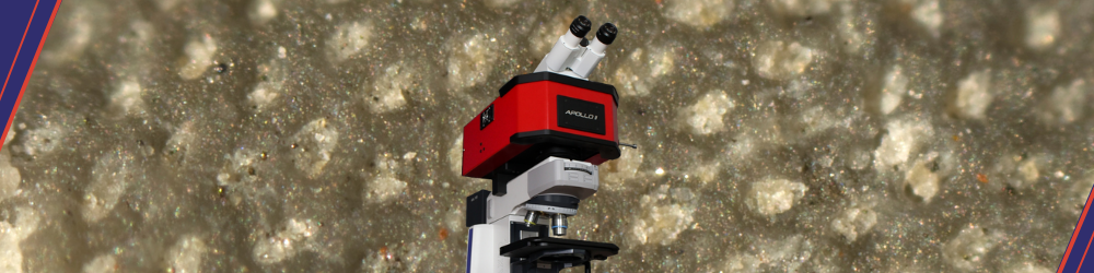
Distinguishing true microfossils from mineral mimics demands precise, multi-layered verification. By combining chemical, spatial, and thermal analysis, scientists can undertake confident identification. CRAIC Technologies' Apollo M™ Raman Microspectrometer integrates confocal imaging with high-sensitivity spectral acquisition, enabling detailed characterisation of carbon at the microscale. Designed for complex geological samples, it establishes the clarity to resolve ambiguous microfossil-like structures. Speak with CRAIC Technologies today to learn more about our Apollo M™ Raman Microspectrometer and how it can improve the accuracy and reliability of your microfossil analysis.
References
- Brasier M, Grassineau N, Green O, et al. Questioning the evidence for Earth’s oldest fossils. Nature. 2002;416:76-81. doi:10.1038/416076a.
- Emry J, Marshall A, Marshall C. Multiple Generations of Carbon in the Apex Chert and Implications for Preservation of Microfossils. Astrobiology. 2012;12(2):160-166. doi:10.1089/ast.2011.0729.
- Bost N, Foucher F, Guimbretière G, et al. “Petrochemical and Mineralogical Applications of Raman Mapping”. Raman Spectroscopy and Applications. Edited by Khan Maaz. InTechOpen. 2017. doi:10.5772/65112.
- Bower DM, Fries MD, Kater L, et al. Micro Raman spectroscopy of carbonaceous material in microfossils and meteorites: improving a method for life detection. Astrobiology. 2013;13(1):103-113. doi:10.1089/ast.2012.0865.
- Bower DM, Fries MD, Green OR, et al. Raman Imaging Spectroscopy of a Putative Microfossil from the 〜3.46 Ga Apex Chart: Insights from Quartz Grain Orientation. Astrobiology. 2016;16(2):169-180. doi:10.1089/ast.2014.1207.