
Using Microspectroscopy to Decode the Structural Color of Butterfly Wings
A butterfly wing may appear uniformly colored to the eye, but its optical behavior can vary considerably across microscopic distances. Individual scales contain intricate structures that manipulate light through interference, diffraction, and scattering, producing structural colors without relying solely on pigments. Since relatively small differences in these structures can alter the resulting spectrum, averaging measurements across a large area risks masking important variations. Microspectroscopy avoids this limitation by measuring the individual scales and ridges that produce them.
The Physics of Structural Color at the Microscale
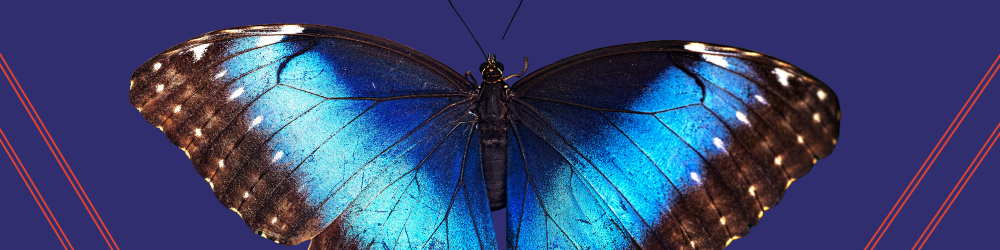
Among nature’s most recognizable examples, Morpho butterflies display an intense metallic blue despite lacking a corresponding blue pigment. Their wing scales contain highly organized chitin ridges with stacked, fir-tree-shaped lamellae. Light reflected from successive layers interferes constructively at selected wavelengths and destructively at others, generating the characteristic blue appearance.
Periodic geometry adds complexity to the butterfly's color response. Ridge spacing, microribs, crossribs, lamellar thickness, and refractive-index differences can alter wavelength selection, spectral intensity, and viewing-angle behavior. Consequently, a small variation in nanoscale geometry may produce a measurable change in color.
Natural chitin structures can also function as photonic crystals. Periodic variations in refractive index regulate how particular wavelengths propagate through the wing-scale architecture, with some structures producing photonic bandgap effects. Such biological systems act as useful models for engineering:
- Anti-reflective coatings for lenses and optical components
- Reflective displays that consume little energy when maintaining an image
- Security features with angle-dependent optical signatures
- Structural colors resistant to chemical fading
- Optical sensors that respond to changes in local refractive index
Individual butterfly scales may measure approximately 50 to 100 µm across, yet their optically active surface structures extend into the sub-micron range. Measuring an entire wing can merge responses from different scales, orientations, pigments, and structural regions, and so obscure localized variations in optical behavior. Microspectroscopy addresses this analytical issue by restricting spectral acquisition to carefully selected microscopic areas.
Decoding Butterfly Structural Color via Microspectroscopy
Microspectroscopy combines optical microscopy with sensitive spectroscopy, allowing a researcher to see a microscopic feature and measure its spectral response from the same region. A single scale can be isolated from its surroundings, and smaller sampling apertures can target localized ridge structures without incorporating signals from neighboring wing areas.
Micro-reflectance spectroscopy then provides a direct method for examining wavelength-selective behavior. Across ultraviolet (UV), visible, and near-infrared (NIR) wavelengths, reflectance spectra reveal maxima and minima produced by constructive and destructive interference.
Researchers can compare the reflectance spectra from individual wing scales or selected microscopic regions with structural measurements and optical models to investigate several parameters. These include:
- Ridge and lamellar spacing
- Multilayer thickness
- Refractive-index contrast
- Periodicity and structural organization
- Local variations across a single scale
Angle-resolved microspectroscopy extends this analysis by examining how spectral behavior changes with illumination or observation angle. Measuring spectral peaks at different angles quantifies iridescence and helps establish how a particular scale architecture directs reflected light.
Polarization analysis can further characterize the optical response of anisotropic wing-scale structures. Anisotropic ridges and asymmetric nanostructures may interact differently with specific polarization states, making polarization-resolved measurements useful for studying directional photonic effects and informing polarization-sensitive optical materials.
Chemical coloration must also be considered. Butterfly scales may contain absorbers such as melanin or pterin pigments alongside light-manipulating chitin structures. Combining micro-reflection, micro-transmission, absorbance, and photoluminescence measurements helps researchers distinguish structural interference from chemical absorption or emission. Microspectroscopy can thus provide a more complete account of how morphology and chemistry contribute to the observed color of the wing.
Microspectroscopy for Advanced Structural Color Analysis
Moving from biological observation to biomimetic engineering calls for spectral data linked to precise microscopic locations. Specialists developing synthetic photonic crystals, patterned optical coatings, structural-color materials, or refractive-index sensors need to determine which physical features generate a particular optical response.
The 2030PV PRO™ Microspectrometer from CRAIC Technologies is designed for localized optical characterization across a broad spectral range. Relevant capabilities include:
- Deep UV to NIR analysis- Measurements from 200 to 2,500 nm support transmission, absorbance, reflectance, and polarization studies across multiple spectral regions
- Controlled microscale sampling- Variable, NIST-traceable micro-apertures enable researchers to isolate selected areas of butterfly scales, photonic crystals, and other heterogeneous specimens
- Sensitive spectral detection- Specialized high-sensitivity detectors support strong signal-to-noise performance when working with delicate or low-reflectance microscopic samples
- Digital sample visualization- High-resolution imaging in UV, visible, and NIR light helps position the measurement aperture over a defined region of interest
Such capabilities are extremely valuable if optical properties vary significantly across a specimen. Scientists can correlate visual morphology with localized spectra, compare neighboring structures, and investigate spectral changes without relying on measurements averaged across large sample areas.
From Butterfly Wings to Biomimetic Materials
Through microspectroscopy, researchers can examine how butterfly-scale architecture manipulates light and apply these observations to the development of engineered photonic materials. CRAIC Technologies' 2030PV PRO™ Microspectrometer supports their work with microscale sampling, broad 200 to 2,500 nm spectral coverage, sensitive detection, and high-resolution imaging. For scientists and engineers studying structural color, optical nanostructures, or biomimetic materials, CRAIC Technologies offers analytical platforms built for microscale characterization. Contact CRAIC Technologies to identify the right microspectroscopy solution for your research.
References
- Roy Sambles J & Vukusic P. Photonic structures in biology. Nature. 2003;424:852-855. doi:10.1038/nature01941.
- Kinoshita S & Yoshioka S. Structural Colors in Nature: The Role of Regularity and Irregularity in the Structure. ChemPhysChem. 2005;6(8):1442-1459. doi:10.1002/cphc.200500007.
- Macedonia J.M, Morehouse N, Rutowski R L, et al. Pterin pigments amplify iridescent ultraviolet signal in males of the orange sulphur butterfly, Colias eurytheme. Proceedings of the Royal Society B. 2005;272(1578):2329-2335. doi:10.1098/rspb.2005.3216.
- Morehouse N, Rutowski R, & Vukusic P. Pterin pigment granules are responsible for both broadband light scattering and wavelength selective absorption in the wing scales of pierid butterflies. Proceedings of the Royal Society B: Biological Sciences. 2007;274(1608):359-366. doi:10.1098/rpsb.2006.3730
- Counterman B, Day C, Hanly J, et al. Convergent Evolution of Broadband Reflectors Underlies Metallic Coloration in Butterflies. Frontiers in Ecology and Evolution. 2020;8:206. doi:10.3389/fevo.2020.00206.
