
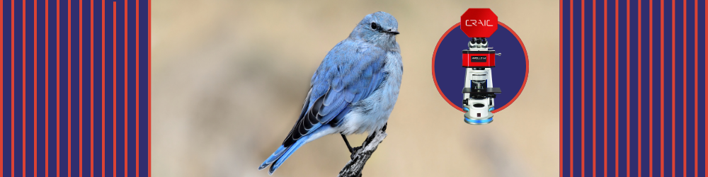
Blue Without Pigment: How Microspectroscopy Proves Bluebirds Aren't Actually Blue
A bluebird appears to offer straightforward evidence of blue pigment, yet its brilliant plumage tells a more complex optical story. Avian reds, yellows, browns, and blacks commonly arise from pigments such as carotenoids and melanins, but true blue pigments are virtually absent from bird feathers. Bluebirds achieve their color through structural coloration, where nanoscale architecture manipulates visible light. Microspectroscopy reveals exactly how that physical mechanism works.
The Physics of Feather Architecture: Coherent Scattering
Beneath the surface of a bluebird feather, individual barbs contain several optically distinct regions. An outer keratin cortex surrounds a spongy medullary layer composed of beta-keratin and nanoscale air spaces. Deeper within the barb lies melanin, which provides a dark optical backing. Far from being decorative anatomy, that layered arrangement determines which wavelengths return to an observer and which disappear inside the feather.
Historically, blue plumage was often explained through Rayleigh or Tyndall scattering, phenomena in which small particles scatter shorter wavelengths more efficiently than longer ones. However, detailed studies of avian coloration have revealed a more specific mechanism: coherent scattering. Within the quasi-ordered keratin-air matrix, nanometer-scale spacing causes scattered light waves at selected wavelengths to reinforce one another through constructive interference. The effect is strongest at blue wavelengths, around 400-475 nm, giving the feather its characteristic blue appearance.
Longer wavelengths behave differently. Much of that light travels through the spongy layer of the feather barb and reaches the underlying melanin, which absorbs it and suppresses broadband backscatter. By removing unwanted wavelengths from the returning signal, melanin increases the saturation of the blue produced by the nanostructure. Consequently, the final color of the feather emerges from an interaction between nanoscale geometry, refractive-index contrast, interference, and absorption.
How Microspectroscopy Exposes the Optical Mechanism of Bluebird Feathers
Reflectance microspectroscopy provides direct spectral evidence from an individual feather barb. Under reflected illumination, a microspectrometer can record a pronounced peak around the blue or violet region, often near 450 nm. Because the instrument targets a microscopic area, researchers can correlate that spectral feature with the precise part of the barb responsible for the observed color.
Transmission microspectroscopy can be used as a complementary test. When light passes through the feather, instead of reflecting from its surface, the strong blue signal becomes much less prominent, while the light-absorbing effect of the underlying melanin becomes clearer. That contrast is important since chemical pigments and structural colors interact with light through different mechanisms. Pigments selectively absorb wavelengths according to molecular electronic transitions, whereas structural coloration arises from the scattering and interference of light caused by physical architecture. Comparing these spectral responses helps distinguish color generated by feather nanostructure from color produced by light-absorbing pigments.
Refractive-index matching can also be applied to test the structural origin of a feather's blue coloring. Air has a refractive index near 1.0, while the refractive index of beta-keratin is approximately 1.55. Introducing a suitable index-matching fluid into the nanoscale air spaces within the feather barb reduces the optical difference between neighbouring regions. With these interfaces suppressed, coherent scattering weakens dramatically and the characteristic blue reflectance can collapse. As the fluid evaporates and air returns, the spectral peak reappears.
Polarized microspectroscopy offers further information about the optical organization of feather nanostructure. Bluebird feather nanostructures are quasi-ordered and largely isotropic, producing non-iridescent coloration that remains comparatively stable across viewing orientations. Highly ordered multilayer structures, such as those found in many iridescent peacock feathers, show much stronger directional and polarization effects. Spatial spectral mapping can distinguish such variations across microscopic regions and relate optical response directly to local feather architecture.
Biomimicry: Translating Feather Optics into Materials
Beyond evolutionary biology, avian coloration offers practical design principles for material scientists and optical engineers. Nanoscale control over spacing and refractive index can generate structural hues without relying on conventional colorants. Researchers are applying comparable photonic principles to fade-resistant coatings, authentication features for banknotes and branded products, low-energy reflective displays, and wavelength-selective optical coatings.
Reflectance microspectroscopy helps characterize the optical properties of engineered photonic materials and nanostructures. Key measurements used to evaluate their optical response include:
- Peak reflectance wavelength and intensity
- Full width at half maximum (FWHM) bandwidth
- Spatial variation across patterned surfaces
- Angular and polarization-dependent spectral response
The data ascertained from reflectance microspectroscopy measurements can reveal which aspects of nanoscale architecture are responsible for specific colors and optical behaviors in bio-inspired photonic systems.
Revealing Nanoscale Optics with CRAIC Technologies
Blue feathers in nature demonstrate why optical appearance cannot always be explained by chemistry alone. CRAIC Technologies provides microspectroscopy systems for investigating such microscopic optical behavior, including the 2030PV PRO™ microspectrophotometer. This instrument supports reflectance, absorbance, fluorescence, and polarization measurements from microscopic sample areas, with spectral coverage extending from the deep ultraviolet through visible wavelengths into the near-infrared. Integrated digital imaging and automated spectral acquisition also help researchers target precise regions efficiently. Contact CRAIC Technologies today for additional information about our microspectroscopy solutions that enable the characterization of structural color, photonic materials, coatings, and other microscale optical technologies.
References
- Dyck J, Prum R, Torres R, et al. Coherent light scattering by blue feather barbs. Nature. 1998;396:28-29. doi:10.1038/23838.
- Morehouse N, Shawkey M, & Vukusic P. A protean palette: colour materials and mixing in birds and butterflies. Journal of Royal Society Interface. 2009;6(2):S221-S231. doi:10.1098/rsif.2008.0459.focus.
- Clarke J, Eliason C, & Kane S. Wrinkle nanostructures generate a novel form of blue structural color in great argus flight feathers. iScience. 2022;26(1):105912. doi:10.1015/j.isci.2022.105912.
